|
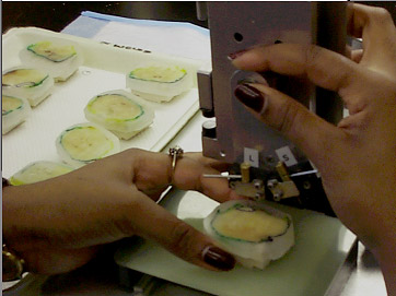
| A Prostate Specimen |
 |
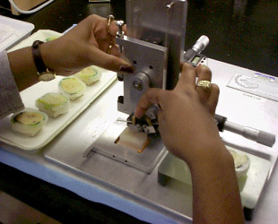
| Blocks |
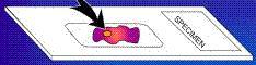 |
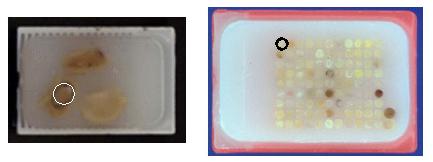
| A slide. The yellow
circle represents a tissueDiagnosis. There may be several
tissueDiagnosises per slide, and each is labelled, given a
letter-designation (a,b,c,etc), and given a diagnosis (normal, cancer,
etc) |